-
出版商:
Springer
-
出版日期:
2026-08-07
-
售價:
$6,640
-
貴賓價:
9.5 折
$6,308
-
語言:
英文
-
頁數:
255
-
裝訂:
Quality Paper - also called trade paper
-
ISBN:
3031931246
-
ISBN-13:
9783031931246
-
相關分類:
理工類
商品描述
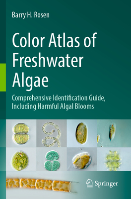
Microscopic observations of the inhabitants in aquatic environments reveal the complex array of organisms that are woven together into a tapestry that is informative, ornate, colorful, and artistic. The diversity of life, especially among the photosynthetic organisms called algae, displays a unique suite of patterns and structures that allows the names to be assigned. This book compiles microphotographs from many groups and hundreds of genera to illustrate the beauty of algae and assist in their identification. The images in this guide were initially started as an academic, hands-on teaching guide rather than a digital version. This first edition organizes and arrays the images in taxonomic groups that can be used to instruct a new generation of phycologists, those that study algae. The identification and classification of algae from Linnaeus in 1767, through the 19th and 20th centuries, mostly relied on pen and ink line drawings, carefully constructed to illustrate morphological features that separated species, and these are still a valuable resource. Both scanning electron microscopy (SEM) and transmission electron microscopy (TEM) brought significantly higher resolution than light microscopy, illustrating the external and internal structures through black and white imagery. As digital photography improved for the light microscope in the past several years, color images of the key features that distinguish one organism from another can be generated.
商品描述(中文翻譯)
對水生環境中生物的顯微觀察,揭示了各種生物彼此交織而成的複雜組合,宛如一幅兼具資訊性、華麗性、色彩與藝術性的織錦。生命的多樣性,尤其是被稱為藻類的光合生物,展現出一組獨特的圖樣與結構,使我們得以為其鑑定與命名。本書彙整了來自許多類群及數百個屬的顯微攝影影像,用以呈現藻類之美,並協助進行鑑定。
本指南中的影像最初是為了製作學術性的實作教學指南而開始蒐集,而非作為數位版本使用。本第一版依照分類群組整理並編排這些影像,可用於教導新一代的藻類學家(研究藻類的學者)。自 Linnaeus 於 1767 年開始,歷經 19 世紀與 20 世紀,藻類的鑑定與分類主要依賴鋼筆與墨水繪製的線條圖;這些圖繪經過精心製作,用來呈現區分不同物種的形態特徵,至今仍是相當寶貴的資源。
掃描式電子顯微鏡(scanning electron microscopy, SEM)與穿透式電子顯微鏡(transmission electron microscopy, TEM)所提供的解析度,顯著高於光學顯微鏡,並透過黑白影像呈現生物的外部與內部結構。過去數年間,隨著光學顯微鏡的數位攝影技術日益進步,我們已能產生呈現關鍵特徵的彩色影像,藉此區分不同生物。
作者簡介
Dr. Rosen has been studying algae for more than 50 years, beginning at the University of Connecticut. He is a world-class scholar and has taught algae workshops and courses around the country and online. His focus on cyanobacteria and harmful algal blooms has led to many invited speeches and media events. The advent of digital microscopic photography has allowed him to produce unique images of algae.
作者簡介(中文翻譯)
Rosen 博士研究藻類已有 50 多年的歷史,最初是在 University of Connecticut 開始這項研究。他是世界級的學者,曾在全美各地及線上教授藻類工作坊與課程。他對藍綠藻(cyanobacteria)和有害藻華(harmful algal blooms)的研究專長,促成許多受邀演講與媒體活動。數位顯微攝影技術的問世,也讓他得以創作出獨特的藻類影像。